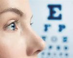

- What Is Keratoconus and How It Affects Vision
- Causes and Risk Factors Behind Keratoconus
- Recognizing Symptoms of Keratoconus Early
- The Diagnosis Process: How Experts Identify Keratoconus
- Treatment Options and Latest Advances in Managing Keratoconus
- Personal Stories and Expert Insights on Living with Keratoconus
- Where to Find Support and Services for Keratoconus - Eye Docs
What Is Keratoconus and How It Affects Vision
Keratoconus is a progressive eye condition where the normally round cornea thins and begins to bulge into a cone-like shape. This irregular shape causes distorted vision and can severely affect daily life if not properly managed. Unlike common refractive errors like nearsightedness or farsightedness, keratoconus alters the fundamental structure of the cornea, leading to visual disturbances such as blurred vision, glare, and sensitivity to light.
The cornea is essential in focusing light onto the retina, and when its shape changes unpredictably, focusing becomes impaired. This can make tasks like reading, driving at night, or using screens frustrating and challenging. Understanding keratoconus and treatment options is critical to preserving vision and maintaining a good quality of life.
How Keratoconus Progresses Over Time
In most cases, keratoconus begins during the teenage years or early twenties and can progress gradually over a decade or more. The rate of progression varies significantly between individuals. Some may experience slow changes that stabilize in middle age, while others see rapid deterioration that requires early intervention.
Because keratoconus develops over time, regular eye exams are vital for early detection and to monitor the condition closely. Early diagnosis opens the door to a wider range of treatment options that can slow or even halt progression.
Causes and Risk Factors Behind Keratoconus
Despite ongoing research, the exact cause of keratoconus remains not fully understood. However, experts believe it results from a combination of genetic predisposition and environmental factors. For example, there is often a family history of keratoconus among patients, suggesting genetic links.
Additional factors contributing to keratoconus development include:
- Eye Rubbing: Persistent and vigorous eye rubbing can weaken the corneal tissue, accelerating thinning.
- Allergies and Inflammation: Chronic eye irritation caused by allergies may increase risk by promoting eye rubbing and inflammation.
- Other Medical Conditions: Some connective tissue disorders have been linked to keratoconus.
Understanding these factors can help in prevention and management strategies. For example, minimizing eye rubbing and controlling allergic symptoms can reduce keratoconus risk or progression.
Recognizing Symptoms of Keratoconus Early
One of the biggest challenges with keratoconus is its subtle onset. Early symptoms can mimic common vision problems, which is why many patients initially mistake them for routine refractive errors. Key symptoms to watch for include:
- Blurred or distorted vision that cannot be fully corrected with glasses
- Increased sensitivity to bright lights and glare
- Frequent changes in eyeglass prescriptions
- Difficulty seeing clearly at night
- Double vision or ghost images in one eye
If you notice any of these signs, especially if they worsen quickly, it’s important to seek professional eye care for thorough evaluation.
Case Highlight: Early Detection Made a Difference
Consider the story of Sarah, a 19-year-old college student who began experiencing blurry vision and frequent prescription changes. Initially, she thought it was normal eye strain, but after a routine checkup, she was diagnosed with keratoconus. Because her condition was caught early, Sarah was able to undergo corneal cross-linking treatment, which helped stabilize her cornea and preserve her vision. Her experience underscores the importance of early recognition and proactive treatment.
The Diagnosis Process: How Experts Identify Keratoconus
Diagnosing keratoconus involves more than just a standard eye exam. Specialists use advanced imaging technology to map the shape and thickness of the cornea with precision. Common diagnostic tools include:
- Corneal Topography: Produces a detailed map of the corneal surface, highlighting irregularities.
- Pachymetry: Measures corneal thickness, a key indicator in keratoconus.
- Slit-Lamp Examination: Allows the eye doctor to inspect corneal structure and signs of thinning or scarring.
These tests enable a comprehensive assessment, guiding the selection of appropriate treatment tailored to the severity of the condition.
Treatment Options and Latest Advances in Managing Keratoconus
Management of keratoconus has evolved significantly in recent years. Treatment options depend on the stage of the disease and how much the vision is affected.
Non-Surgical Interventions
In mild to moderate keratoconus, vision can often be improved with specially designed contact lenses. These include:
- Rigid Gas Permeable (RGP) Lenses: These lenses create a smooth optical surface, improving vision.
- Scleral Lenses: Larger lenses that vault over the cornea, providing comfort and clear vision even in advanced cases.
- Hybrid Lenses: Combining a hard center with a soft outer ring, these lenses offer comfort and vision correction.
These options can significantly improve quality of life but require fitting by specialists familiar with keratoconus.
Advanced Treatments
For progressive cases, newer treatments aim to strengthen the cornea and prevent further deterioration:
- Corneal Cross-Linking (CXL): This minimally invasive procedure uses ultraviolet light and riboflavin drops to strengthen corneal collagen fibers, halting progression.
- Intacs: Small ring implants inserted into the cornea to reshape it and improve vision.
- Corneal Transplant: Reserved for severe cases where scarring or thinning has severely impaired vision, a transplant replaces the damaged cornea.
Emerging research continues to improve these approaches, with ongoing innovations enhancing safety and effectiveness.
Personal Stories and Expert Insights on Living with Keratoconus
Living with keratoconus can be challenging, both physically and emotionally. Many patients report anxiety over vision changes and adapting to new treatments. However, expert ophthalmologists emphasize that with timely diagnosis and tailored treatment, most people maintain good functional vision for years.
For example, John, a 34-year-old graphic designer, shared how switching to scleral lenses allowed him to continue his work and hobbies without interruption. He stresses the importance of ongoing monitoring and staying informed about new treatment options.
Professionals recommend regular follow-ups and encourage patients to seek support from eye care communities. This approach not only ensures optimal management but also helps patients feel less isolated in their journey.
Where to Find Support and Services for Keratoconus - Eye Docs
For those seeking comprehensive keratoconus care, Eye Docs offers expert consultations, fitting services for specialized contact lenses, and access to the latest treatment technologies. Whether you need detailed diagnosis, personalized treatment plans, or ongoing management support, Eye Docs provides reliable and patient-focused solutions.
Visiting Eye Docs ensures that you are guided by experienced professionals who understand the complexities of keratoconus and can help you navigate treatment choices tailored to your needs.